X-rays, a form of electromagnetic radiation, have revolutionized the field of medicine since their discovery by Wilhelm Conrad Roentgen in 1895. As an X-ray supplier, I've witnessed firsthand how these remarkable rays have become an indispensable tool in modern healthcare. In this blog, I'll explore the diverse uses of X-rays in medicine, highlighting their significance and the types of X-ray machines available to meet various medical needs.
Diagnostic Imaging
One of the primary uses of X-rays in medicine is diagnostic imaging. X-ray machines are capable of producing detailed images of the internal structures of the body, allowing healthcare professionals to detect and diagnose a wide range of conditions.
Bone and Joint Examinations
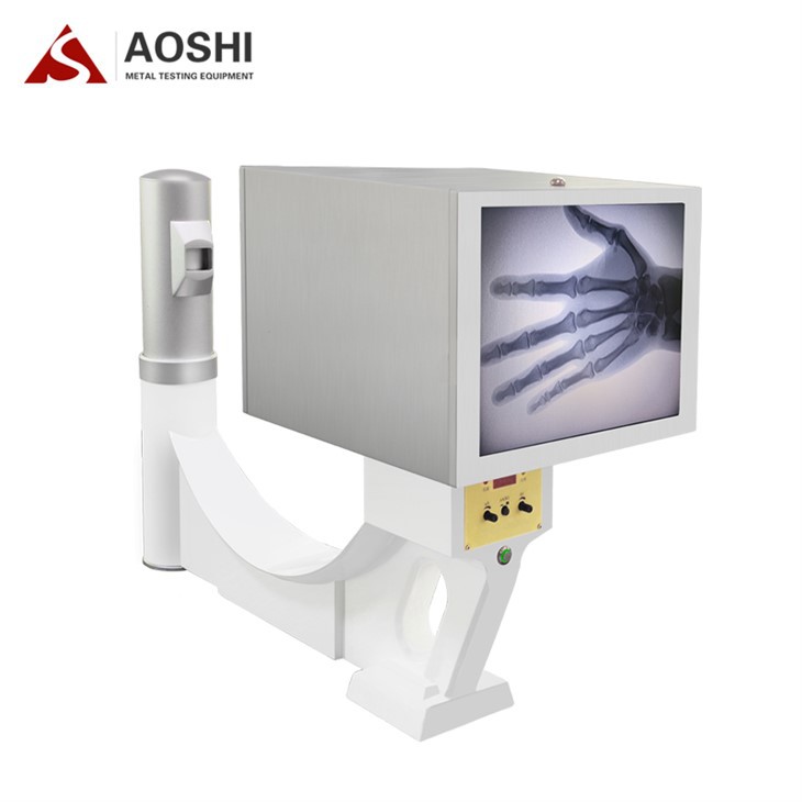
X-rays are commonly used to examine bones and joints, making them invaluable in orthopedics. Orthopedic X-ray Machine can provide clear images of fractures, dislocations, arthritis, and other bone-related conditions. These images help doctors determine the extent of the injury or disease and develop appropriate treatment plans. For example, in the case of a suspected broken bone, an X-ray can quickly confirm the diagnosis and show the location and severity of the fracture, guiding the decision on whether to use a cast, splint, or surgery.
Dental X-rays
In dentistry, X-rays play a crucial role in detecting dental problems that are not visible to the naked eye. Dental X-rays can reveal cavities between teeth, bone loss due to periodontal disease, impacted teeth, and other oral health issues. This information is essential for dentists to provide accurate diagnoses and develop effective treatment strategies, such as fillings, extractions, or orthodontic treatments.

Chest X-rays
Chest X-rays are a common diagnostic tool for evaluating the health of the lungs and heart. They can detect conditions such as pneumonia, lung cancer, tuberculosis, and heart enlargement. By providing a clear image of the chest cavity, chest X-rays help doctors identify abnormalities and determine the appropriate course of treatment. For instance, in the case of a patient with shortness of breath, a chest X-ray can help rule out or confirm a lung infection or other respiratory conditions.
Fluoroscopy
Fluoroscopy is a real-time imaging technique that uses X-rays to visualize the movement of internal organs and structures. This is particularly useful during certain medical procedures, such as guiding the placement of catheters, needles, or other medical devices. For example, during an angiogram, a contrast agent is injected into the blood vessels, and fluoroscopy is used to monitor the flow of the contrast agent and identify any blockages or abnormalities in the blood vessels. Portable X-ray Machine are often used for fluoroscopy in situations where mobility is required, such as in the operating room or at the bedside.
Mammography
Mammography is a specialized form of X-ray imaging used to screen for breast cancer. It involves taking low-dose X-ray images of the breasts to detect early signs of cancer, such as small tumors or calcifications. Mammograms are recommended for women over a certain age or those at high risk of breast cancer. Early detection through mammography can significantly improve the chances of successful treatment and survival. Our company offers advanced mammography systems that provide high-quality images with low radiation exposure, ensuring the safety and comfort of patients.
Computed Tomography (CT) Scans
CT scans combine multiple X-ray images taken from different angles to create detailed cross-sectional images of the body. This technique provides a more comprehensive view of the internal structures compared to traditional X-rays. CT scans are used to diagnose a wide range of conditions, including tumors, internal injuries, and vascular diseases. They are particularly useful for detecting small lesions or abnormalities that may not be visible on a standard X-ray. Microfocal X-ray Machine are often used in CT scanners to provide high-resolution images, especially for small or delicate structures.
Therapeutic Applications
In addition to diagnostic uses, X-rays also have therapeutic applications in medicine. Radiation therapy, also known as radiotherapy, uses high-energy X-rays or other forms of radiation to kill cancer cells and shrink tumors. This treatment is often used in combination with surgery, chemotherapy, or other cancer treatments. Radiation therapy can be delivered externally using a machine called a linear accelerator or internally using radioactive sources placed near the tumor. Our X-ray machines are designed to meet the strict requirements of radiation therapy, ensuring accurate and precise delivery of radiation to the target area while minimizing damage to surrounding healthy tissues.
Safety Considerations
While X-rays are a powerful diagnostic and therapeutic tool, it's important to use them safely to minimize the risk of radiation exposure. Healthcare providers follow strict guidelines and protocols to ensure that the benefits of X-ray imaging outweigh the potential risks. This includes using the lowest possible radiation dose necessary to obtain the required images, shielding patients from unnecessary radiation, and limiting the frequency of X-ray examinations. As an X-ray supplier, we are committed to providing high-quality X-ray machines that are designed with safety features to protect patients and healthcare workers.
Conclusion
X-rays have had a profound impact on the field of medicine, enabling healthcare professionals to diagnose and treat a wide range of conditions more effectively. From bone and joint examinations to cancer treatment, X-rays play a vital role in modern healthcare. As an X-ray supplier, we are dedicated to providing innovative and reliable X-ray solutions to meet the evolving needs of the medical community. If you are interested in learning more about our X-ray machines or have any questions about their applications, please don't hesitate to contact us for a procurement discussion. We look forward to working with you to improve patient care and outcomes.
References
- Bushberg, J. T., Seibert, J. A., Leidholdt, E. M., & Boone, J. M. (2012). The essential physics of medical imaging. Lippincott Williams & Wilkins.
- Hall, E. J., & Giaccia, A. J. (2012). Radiobiology for the radiologist. Lippincott Williams & Wilkins.
- Hendee, W. R., & Ritenour, E. R. (2002). Medical imaging physics. Wiley-Liss.