As a supplier of Medical X-ray Machines, I've witnessed firsthand the remarkable evolution of this technology and the diverse range of machines available in the market. In this blog, I'll delve into the different types of medical X-ray machines, their unique features, and applications.
1. Conventional X-ray Machines
Conventional X-ray machines are the most basic and widely used type in medical facilities. These machines work by passing a controlled amount of X-ray radiation through the body part being examined onto a detector. The resulting image shows the internal structures based on how different tissues absorb X-rays. Dense tissues like bones appear white, while softer tissues such as muscles and organs are gray, and air-filled spaces are black.
Fixed Installation: Most conventional X-ray machines are fixed in a dedicated radiology room. They are large and stationary, designed to provide high - quality images for a variety of examinations. For example, in a hospital's radiology department, a fixed conventional X-ray machine can be used for chest X-rays to detect conditions like pneumonia, broken ribs, or lung tumors. It can also be used for limb X-rays to diagnose fractures.
General Purpose: These machines are suitable for a wide range of general diagnostic purposes. They are cost - effective and reliable, making them a staple in many healthcare settings, from small clinics to large hospitals. However, they have limitations in terms of mobility and the ability to provide real - time imaging.
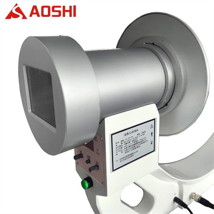
2. Portable X-ray Machines
Portable X-ray machines offer a high degree of flexibility and convenience. As the name suggests, they can be easily moved from one location to another within a healthcare facility or even taken outside the hospital, such as to a patient's bedside in an intensive care unit or to a remote area for on - site medical examinations.
Mobility Advantage: Portable X-ray machines are ideal for situations where moving the patient is difficult or not advisable. For instance, in a trauma unit, a patient with multiple injuries may not be stable enough to be transported to a radiology department. A portable X-ray machine can be brought directly to the patient's bed to quickly assess fractures or internal injuries. You can learn more about Portable X-ray Machine on our website.
Battery - Powered: Many portable X-ray machines are battery - powered, which eliminates the need for a continuous power supply. This feature makes them particularly useful in emergency situations or in areas with unreliable electricity. They are also relatively lightweight and easy to operate, allowing healthcare providers to obtain quick X-ray images without the need for complex setup.
3. Fluoroscopy Machines
Fluoroscopy machines provide real - time, dynamic imaging of the internal organs and structures. They work by continuously emitting X-rays while the patient is being examined, allowing the healthcare provider to observe the movement and function of organs in real time.
Dynamic Imaging: Fluoroscopy is commonly used in procedures such as barium swallow studies, where the patient drinks a contrast agent, and the movement of the agent through the digestive tract is observed. It is also used in interventional radiology procedures, such as guiding the placement of catheters or stents. The real - time imaging capability of fluoroscopy machines helps doctors make more accurate diagnoses and perform procedures with greater precision.
High - Dose Radiation: However, one of the drawbacks of fluoroscopy is that it exposes the patient to a relatively higher dose of radiation compared to conventional X-ray machines. Therefore, it is important to use fluoroscopy only when necessary and to take appropriate radiation protection measures.
4. Mammography Machines
Mammography machines are specifically designed for breast imaging. They use low - dose X-rays to detect early signs of breast cancer, such as microcalcifications or masses that may not be palpable during a physical examination.
Specialized Design: Mammography machines have a unique design that allows for the compression of the breast tissue to obtain clear and detailed images. The compression helps to spread out the breast tissue, reducing the amount of overlapping and improving the visibility of any abnormalities. There are two main types of mammography: film - screen mammography and digital mammography.
Digital Mammography: Digital mammography has become increasingly popular in recent years due to its ability to produce high - quality images that can be easily stored, manipulated, and transmitted. It also allows for better visualization of breast tissue in women with dense breasts. Early detection of breast cancer through mammography can significantly improve the chances of successful treatment.
5. Dental X-ray Machines
Dental X-ray machines are used in dentistry to diagnose a variety of oral conditions, including tooth decay, gum disease, and impacted teeth. There are several types of dental X-ray machines, each with its own specific application.
Intraoral X-ray Machines: Intraoral X-ray machines are used to take images of individual teeth or small groups of teeth. The X-ray film or digital sensor is placed inside the patient's mouth, and the X-ray beam is directed at the area of interest. These machines are commonly used for routine dental check - ups and to detect cavities or other dental problems.
Panoramic X-ray Machines: Panoramic X-ray machines provide a wide - view image of the entire mouth, including the teeth, jaws, and surrounding structures. They are useful for diagnosing conditions such as wisdom tooth impaction, jaw fractures, and periodontal disease. Dental X-ray machines are designed to minimize the radiation exposure to the patient while providing clear and accurate images.
6. Computed Tomography (CT) Scanners
CT scanners are advanced X-ray machines that produce cross - sectional images of the body. They work by rotating an X-ray tube and detector around the patient, taking multiple X-ray images from different angles. A computer then processes these images to create detailed, three - dimensional images of the internal organs and structures.
High - Resolution Imaging: CT scanners offer high - resolution imaging that can detect small lesions, tumors, and other abnormalities that may not be visible on conventional X-ray images. They are commonly used in the diagnosis of a wide range of conditions, including head injuries, lung diseases, and abdominal disorders.
Contrast - Enhanced CT: In some cases, a contrast agent may be administered to the patient before a CT scan to improve the visibility of certain organs or blood vessels. However, CT scans expose the patient to a relatively higher dose of radiation compared to other X-ray modalities, so they are typically used when the benefits of the examination outweigh the potential risks.
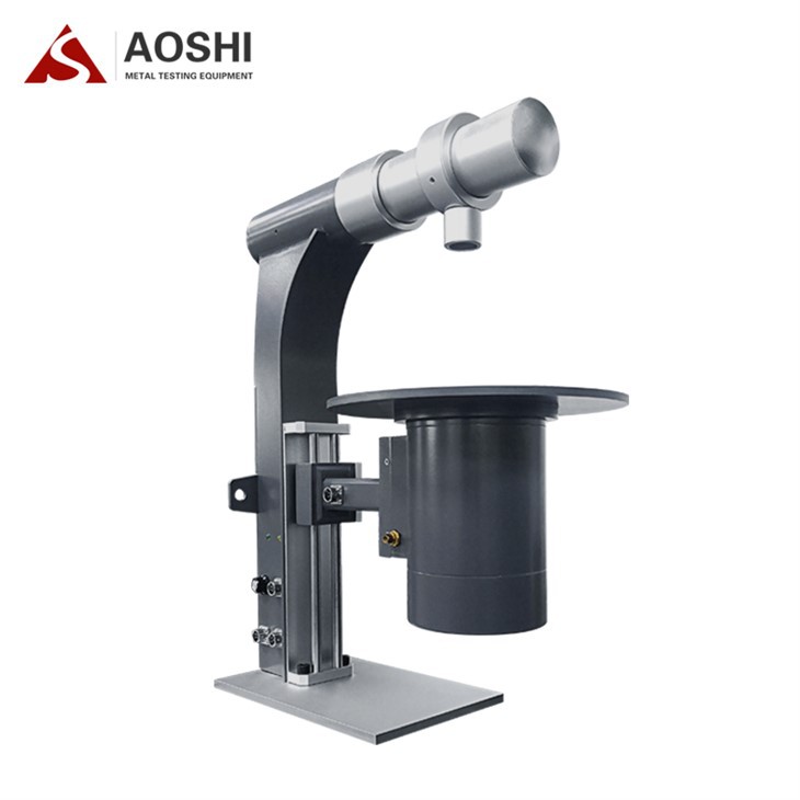
7. Industrial X-ray Machines (Related in some aspects)
Although not strictly medical X-ray machines, Industrial X-ray Machine are worth mentioning as they share some technological similarities. Industrial X-ray machines are used in various industries, such as manufacturing, aerospace, and electronics, to inspect the internal structure of materials and components for defects or damage.
Non - Destructive Testing: These machines use X-rays to detect flaws, cracks, or inclusions in materials without damaging them. The technology behind industrial X-ray machines, such as the generation and detection of X-rays, is similar to that of medical X-ray machines. However, the applications and safety requirements are different.
Conclusion
The world of medical X-ray machines is vast and diverse, with each type of machine offering unique features and benefits. As a supplier of Medical X-ray Machine, we understand the importance of providing high - quality, reliable, and innovative X-ray solutions to meet the needs of healthcare providers and patients. Whether you are looking for a basic conventional X-ray machine for your clinic or a state - of - the - art CT scanner for a large hospital, we have the expertise and products to help you make the right choice.
If you are interested in learning more about our medical X-ray machines or would like to discuss your specific requirements, please feel free to contact us. We are committed to providing excellent customer service and support throughout the purchasing process and beyond. Let's work together to improve the quality of healthcare through advanced X-ray technology.
References
- Bushberg, J. T., Seibert, J. A., Leidholdt, E. M., & Boone, J. M. (2012). The essential physics of medical imaging. Lippincott Williams & Wilkins.
- Hendee, W. R., & Ritenour, E. R. (2002). Medical imaging physics. Wiley - Liss.
- Fauber, T. L. (2009). Radiographic imaging and exposure. Elsevier Health Sciences.