Fluoroscopy is a real-time imaging technique that uses X-rays to obtain moving images of the internal structures of a patient. It's like a live-action movie of what's going on inside the body, as opposed to a static photo you'd get from a regular X-ray. As an X-ray supplier, I've seen firsthand how crucial fluoroscopy is in a wide range of medical procedures.
How Fluoroscopy Works
Let's start with the basics of how fluoroscopy works. A fluoroscopy machine consists of an X-ray tube that emits X-rays and an image intensifier that captures the X-rays after they pass through the body. The image intensifier converts the X-rays into a visible light image, which is then displayed on a monitor in real-time. This allows doctors to see the movement and function of organs and tissues as they happen.
Think of it as a high-tech window into the body. The X-rays pass through different tissues in the body at different rates. Dense tissues like bones absorb more X-rays and appear white on the monitor, while softer tissues like muscles and organs absorb fewer X-rays and appear in shades of gray. Fluids, like blood or contrast agents, can also be visualized, which is super helpful for many procedures.
Uses in Diagnostic Procedures
One of the most common uses of fluoroscopy is in diagnostic procedures. For example, in a barium swallow test, a patient drinks a liquid containing barium, which is a contrast agent. As the barium moves through the esophagus, stomach, and intestines, the fluoroscopy machine can capture real-time images of the digestive tract. This helps doctors detect problems like ulcers, tumors, or blockages.
In orthopedics, fluoroscopy is used to guide the placement of pins, screws, and plates during fracture repair. Surgeons can use the real-time images to ensure that the hardware is properly positioned, which is crucial for a successful outcome. You can learn more about orthopedic X-ray machines on our website Orthopedic X-ray Machine.

Fluoroscopy is also used to diagnose joint problems. By injecting a contrast agent into a joint and then using fluoroscopy to visualize the joint, doctors can detect issues like cartilage damage, ligament tears, or joint inflammation. This is especially useful for diagnosing problems in the knees, shoulders, and hips. Check out X-ray of Extremities for more information on X-rays for joints and extremities.
Interventional Procedures
In addition to diagnostic uses, fluoroscopy is widely used in interventional procedures. One of the most well-known examples is cardiac catheterization. During this procedure, a thin tube called a catheter is inserted into a blood vessel and guided to the heart. Fluoroscopy is used to visualize the catheter's path and to inject a contrast agent into the blood vessels of the heart. This allows doctors to see the blood flow in the heart and detect blockages or other problems.
Another common interventional use is in the placement of stents. Stents are small, mesh-like tubes that are used to keep blood vessels or other tubular structures open. Fluoroscopy helps doctors guide the stent into the correct position and ensure that it is properly deployed.
Fluoroscopy is also used in pain management procedures. For example, in a nerve block, a local anesthetic is injected near a nerve to block pain signals. Fluoroscopy can be used to guide the needle to the correct location, increasing the accuracy of the injection and reducing the risk of complications.
Benefits of Fluoroscopy
There are several benefits to using fluoroscopy in medical procedures. First and foremost, it provides real-time imaging, which allows doctors to make immediate decisions during a procedure. This can lead to more accurate diagnoses and better treatment outcomes.
Fluoroscopy is also minimally invasive compared to traditional surgical procedures. Many procedures that used to require large incisions can now be performed using small catheters or needles, which reduces the risk of infection, pain, and recovery time for the patient.
Another advantage is that fluoroscopy can be used to guide the placement of medical devices with high precision. This is especially important in procedures where the correct placement of a device is critical for its effectiveness.
Limitations and Risks
Of course, like any medical procedure, fluoroscopy has its limitations and risks. One of the main limitations is the radiation exposure. Since fluoroscopy uses X-rays, patients are exposed to a small amount of radiation. However, the benefits of the procedure usually outweigh the risks, especially when the procedure is necessary for diagnosis or treatment.
Another limitation is that fluoroscopy provides a two-dimensional image, which may not provide a complete view of the three-dimensional structure of the body. In some cases, additional imaging techniques like CT scans or MRI may be needed to get a more detailed picture.
Our Role as an X-ray Supplier
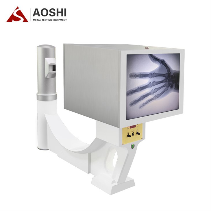
As an X-ray supplier, we play a crucial role in providing high-quality fluoroscopy equipment to medical facilities. Our Medical X-ray Machine is designed to meet the needs of different medical procedures, from simple diagnostic tests to complex interventional procedures.
We offer a range of features and options to ensure that our machines are user-friendly, reliable, and efficient. For example, our machines are equipped with advanced image processing technology to provide clear and detailed images. They also have adjustable settings to optimize the radiation dose for each patient, reducing the risk of unnecessary radiation exposure.
In addition to providing high-quality equipment, we also offer comprehensive after-sales support. Our team of experts is available to provide training, maintenance, and technical support to ensure that our customers can use our machines effectively and safely.
Contact Us for Procurement
If you're a medical facility looking to purchase fluoroscopy equipment or any other X-ray machines, we'd love to hear from you. We understand that choosing the right equipment is an important decision, and we're here to help you make the best choice for your needs.
Whether you're a small clinic or a large hospital, we have the expertise and the products to meet your requirements. Contact us today to start a conversation about how we can provide you with the X-ray equipment you need to improve patient care.
References
- Bushberg, J. T., Seibert, J. A., Leidholdt, E. M., & Boone, J. M. (2012). The essential physics of medical imaging. Lippincott Williams & Wilkins.
- Hall, E. J., & Giaccia, A. J. (2012). Radiobiology for the radiologist. Lippincott Williams & Wilkins.
- Kahn, F. M., & Gibbons, J. P. (2014). Kahn's the physics of radiation therapy. Lippincott Williams & Wilkins.