Hey there! As a supplier of X-ray fluoroscopy equipment, I often get asked about how X-ray fluoroscopy works, especially when it comes to examining the large intestine. So, I thought I'd break it down for you in a way that's easy to understand.
First off, let's talk about what X-ray fluoroscopy is. It's a type of imaging technique that uses X-rays to create real - time moving images of the inside of the body. It's kind of like a live X - ray movie. Instead of just getting a single still image like in a regular X - ray, you can see how organs are functioning and moving.
When it comes to the large intestine, X - ray fluoroscopy can be super useful. The large intestine, also known as the colon, plays a crucial role in our digestive system. It absorbs water from the remaining indigestible food matter and then passes the useless waste material from the body. Problems in the large intestine can range from minor issues like constipation to more serious conditions such as tumors or blockages.
So, how does the whole process of using X - ray fluoroscopy to examine the large intestine actually work?

Preparation
Before the procedure, the patient usually has to do some prep work. This typically involves a special diet for a day or two before the exam. The goal is to clear out the large intestine as much as possible. Sometimes, patients are also given laxatives or enemas to help with this process. This is really important because any remaining feces in the intestine can interfere with the quality of the X - ray images.
Contrast Material
Once the patient is prepped, a contrast material is introduced into the large intestine. There are two main types of contrast: barium sulfate and water - soluble contrast agents. Barium sulfate is a white, chalky substance that shows up very clearly on X - rays. It's usually administered through an enema. The patient lies on a table, and a tube is inserted into the rectum, through which the barium is slowly pumped in. The barium coats the lining of the large intestine, making it easier to see the structure and any potential abnormalities.
Water - soluble contrast agents are used in some cases, especially when there's a risk of the contrast leaking into the abdominal cavity. They are absorbed by the body more quickly and are less likely to cause problems if they do leak.
The X - ray Fluoroscopy Procedure
Now, the patient is ready for the actual X - ray fluoroscopy. They are positioned on an X - ray table, which can be tilted and moved in different directions. The X - ray fluoroscopy machine has an X - ray tube on one side of the table and an image intensifier on the other. The X - ray tube emits a continuous stream of low - dose X - rays through the patient's body. The image intensifier then captures the X - rays that pass through the body and converts them into a visible image on a monitor.
As the contrast material moves through the large intestine, the radiologist can watch the real - time images on the monitor. They can see how the intestine is moving, if there are any blockages, and if the shape and structure of the intestine are normal. The radiologist can also take still images at key moments during the procedure for further analysis.
Different Views
To get a comprehensive view of the large intestine, the radiologist will usually take images from different angles. They might tilt the table to make the contrast material flow in different directions, or ask the patient to move into different positions like lying on their side or standing up. This helps to visualize different parts of the large intestine clearly.
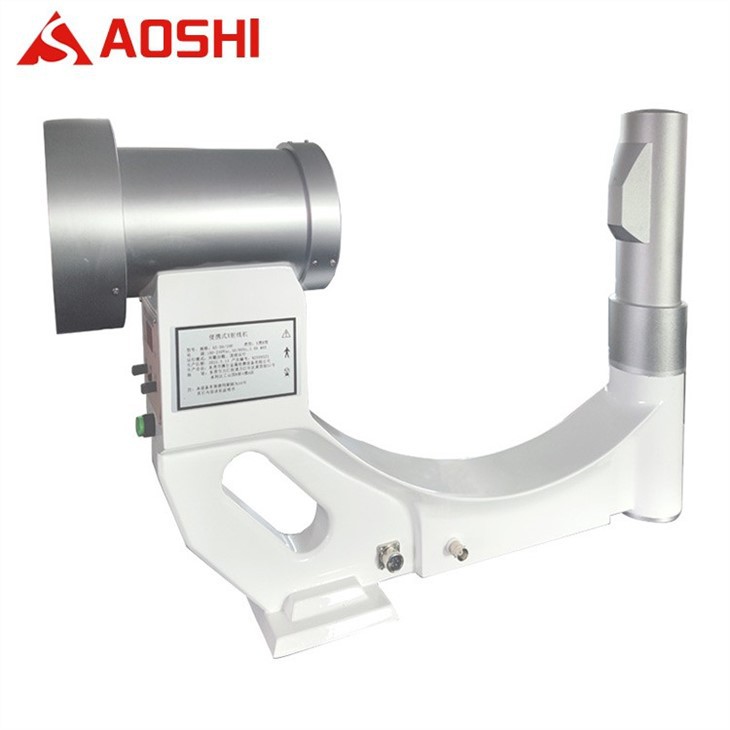
Our X - ray Fluoroscopy Equipment
At our company, we offer a range of high - quality X - ray fluoroscopy equipment. We have Microfocal X-ray Machine, which is great for detailed imaging. It can provide very sharp and clear images, allowing for more accurate diagnosis. Our Medical X-ray Machine is designed specifically for medical applications, including large intestine examinations. It's user - friendly and has advanced features to ensure the best possible results. And if you're looking for something for industrial applications (although not directly related to large intestine exams, but still useful in many other fields), we also have the Industrial X-ray Machine.
Advantages of X - ray Fluoroscopy for Large Intestine Exams
One of the big advantages of X - ray fluoroscopy for large intestine exams is that it's relatively quick and non - invasive compared to some other imaging methods. The patient doesn't have to undergo a major surgical procedure to get a look inside the intestine. It also provides real - time information, which is really helpful for the radiologist to make an accurate diagnosis right away.
Another advantage is that it can detect a wide range of problems. Whether it's a small polyp, a large tumor, or a blockage, X - ray fluoroscopy can often spot these issues. And the use of contrast material makes it even more effective at highlighting the structures in the large intestine.
Limitations
Of course, like any medical procedure, X - ray fluoroscopy has its limitations. One concern is the exposure to radiation. Although the dose is relatively low, repeated exposure to X - rays can increase the risk of certain health problems, such as cancer. That's why it's important to only use this procedure when it's really necessary.
Also, X - ray fluoroscopy might not be able to detect very small or early - stage lesions. In some cases, other imaging methods like CT scans or endoscopy might be needed for a more detailed examination.
Final Thoughts and Contact
If you're in the market for X - ray fluoroscopy equipment for large intestine examinations or any other medical applications, we're here to help. Our team of experts can provide you with all the information you need and assist you in choosing the right equipment for your needs. Whether you're a small clinic or a large hospital, we have solutions that can fit your requirements.
Don't hesitate to reach out to us if you're interested in learning more or starting a purchase negotiation. We're committed to providing high - quality products and excellent customer service.
References
- Bushberg, J. T., Seibert, J. A., Leidholdt, E. M., & Boone, J. M. (2012). The essential physics of medical imaging. Lippincott Williams & Wilkins.
- Sutton, D. (2002). Textbook of radiology and imaging. Churchill Livingstone.