Medical X-ray machines have long been a cornerstone in the field of healthcare, aiding in the diagnosis and treatment of various medical conditions. One of the frequently asked questions is whether these machines can be used for real-time imaging. As a supplier of Medical X-ray Machine, I am well - versed in the technology and capabilities of these devices, and I'm here to provide an in - depth analysis of this topic.
Understanding Real - Time Imaging
Real - time imaging refers to the ability to capture and display images instantaneously, allowing medical professionals to observe dynamic processes within the body as they occur. This is particularly useful in procedures such as guiding minimally invasive surgeries, monitoring the movement of organs, and assessing the function of the cardiovascular system.
Traditional X - ray Machines and Their Limitations
Traditional X - ray machines work by passing a controlled amount of X - ray radiation through the body onto a detector, which then produces a static image. These machines are excellent for detecting fractures, evaluating the presence of foreign objects, and identifying certain types of tumors. However, they are not designed for real - time imaging. The process of capturing an X - ray image typically involves a single exposure, and the resulting image is a snapshot of the body at that moment. It does not provide continuous visual information about moving structures or dynamic physiological processes.
Types of X - ray Machines Capable of Real - Time Imaging
Fluoroscopy
Fluoroscopy is a technique that uses X - rays to obtain real - time moving images of the internal structures of a patient. It works by continuously emitting X - rays while the patient is being examined. The X - rays pass through the body and are detected by a special camera or detector, which then converts the X - ray signals into a video image that is displayed on a monitor. Fluoroscopy is widely used in various medical procedures, such as barium studies to examine the digestive tract, cardiac catheterization to visualize the blood vessels of the heart, and orthopedic procedures to guide the placement of pins and screws.
Microfocal X - ray Machine
Microfocal X - ray machines are another type of X - ray device that can provide high - resolution real - time imaging. These machines use a small focal spot to produce sharp and detailed images. They are often used in research, industrial applications, and some specialized medical fields. In the medical context, microfocal X - ray machines can be used to study small anatomical structures or to monitor the growth and development of cells and tissues in real - time.

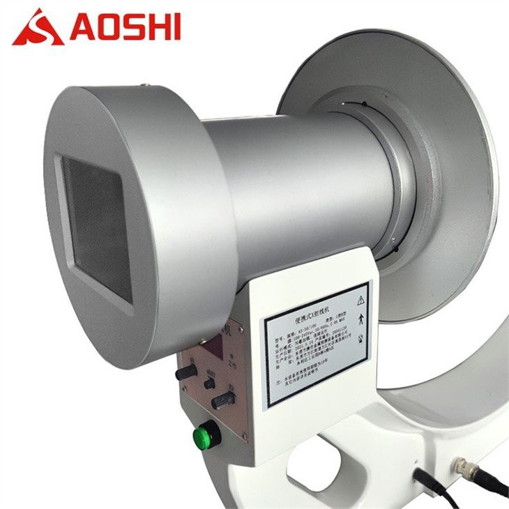
Portable X - ray Machine
Some modern portable X - ray machines are also equipped with real - time imaging capabilities. These machines are designed to be lightweight and easy to move, making them ideal for use in emergency departments, intensive care units, and even in the field. Portable X - ray machines with real - time imaging can quickly provide valuable information about a patient's condition, especially in situations where time is of the essence.
Advantages of Real - Time Imaging with X - ray Machines
Improved Diagnosis
Real - time imaging allows doctors to observe the movement and function of organs and tissues, which can provide more accurate and detailed information for diagnosis. For example, in a fluoroscopic examination of the esophagus, doctors can see how food is being swallowed and whether there are any abnormalities in the movement of the esophageal muscles.
Guided Interventions
During minimally invasive procedures, real - time X - ray imaging provides immediate feedback to the surgeon, allowing for precise placement of instruments and accurate targeting of the affected area. This reduces the risk of complications and improves the success rate of the procedure.
Monitoring Treatment Progress
Real - time imaging can be used to monitor the effectiveness of treatment over time. For example, in patients undergoing chemotherapy or radiation therapy, real - time X - ray imaging can show changes in the size and appearance of tumors, helping doctors adjust the treatment plan as needed.
Challenges and Considerations
Radiation Exposure
One of the main concerns with real - time X - ray imaging is the potential for increased radiation exposure to the patient and medical staff. Since real - time imaging involves continuous or repeated X - ray exposures, it is important to carefully balance the benefits of the imaging with the risks associated with radiation. Medical facilities must follow strict safety protocols to minimize radiation exposure, such as using the lowest possible radiation dose and appropriate shielding.
Image Quality
Achieving high - quality real - time images can be challenging, especially in situations where there is significant movement or when imaging complex anatomical structures. Factors such as patient movement, X - ray scatter, and detector limitations can affect the clarity and accuracy of the images. Advanced imaging techniques and image processing algorithms are often used to improve image quality.
Cost and Availability
Real - time X - ray imaging systems can be expensive to purchase and maintain. Additionally, not all medical facilities may have access to these advanced technologies, especially in resource - limited settings. This can limit the widespread use of real - time X - ray imaging in some regions.
Conclusion
In conclusion, medical X - ray machines can indeed be used for real - time imaging, thanks to technologies such as fluoroscopy, microfocal X - ray machines, and portable X - ray machines with real - time capabilities. Real - time imaging offers significant advantages in terms of diagnosis, treatment guidance, and monitoring, but it also comes with challenges such as radiation exposure, image quality issues, and cost.
As a supplier of Medical X - ray Machine, we are committed to providing high - quality X - ray imaging solutions that meet the diverse needs of our customers. Our products are designed to offer the latest technology in real - time imaging while ensuring patient safety and image quality. If you are interested in learning more about our X - ray machines or are considering purchasing a system for your medical facility, we invite you to contact us for a detailed discussion. We look forward to the opportunity to work with you and help you enhance your diagnostic and treatment capabilities.
References
- Bushberg, J. T., Seibert, J. A., Leidholdt, E. M., & Boone, J. M. (2012). The essential physics of medical imaging. Lippincott Williams & Wilkins.
- Hendee, W. R., & Ritenour, E. R. (2002). Medical imaging physics. Wiley - Liss.
- Fauber, T. L. (2009). Radiographic imaging and exposure. Mosby.